Original

Application of neuroendoscopic surgery in treatment of hypertensive basal ganglia hemorrhage

Z. Zhang, Z. Li [REV NEUROL 2022;75:109-116] PMID: 35880964 DOI: https://doi.org/10.33588/rn.7505.2021445 OPEN ACCESS
Volumen 75 | Number 05 | Nº of views of the article 4.580 | Nº of PDF downloads 90 | Article publication date 01/09/2022
Icono-PDF-OFF Download PDF Castellano Icono-PDF-OFF Download PDF English students-cap copy Acceder a Formación Citation Search in PubMed
Share in: Facebook Twitter
Go to another issue
ABSTRACT Artículo en español English version
INTRODUCTION The influence of Application of neuroendoscopic surgery in treatment of hypertensive basal ganglia hemorrhage remains largely unknown.

AIM To compare the clinical efficacy of minimally invasive neuroendoscopic surgery (NES) and small bone window craniotomy (SBWC) microsurgery on the treatment of patients with hypertensive basal ganglia hemorrhage (HBGH).

PATIENTS AND METHODS The clinical data of 174 HBGH patients treated in our hospital from January 2018 to September 2020 were retrospectively analyzed. They were divided into minimally invasive NES group (n = 90) and SBWC microsurgery group (n = 84). Their operation time, hematoma clearance rate, rebleeding and prognosis were compared.

RESULTS In minimally invasive NES group, the operation time and intraoperative hemostasis time were significantly shorter, and the intraoperative blood loss was significantly less than those in SBWC microsurgery group (p < 0.001). The preoperative Glasgow coma scale (GCS) score was 8.64 ± 1.04 points and 8.68 ± 1.02 points respectively in minimally invasive NES group and SBWC microsurgery group (p > 0.05). At 24 h after operation, the GCS score in minimally invasive NES group rose to 12.89 ± 1.56 points, and it had a significant difference from that in SBWC microsurgery group (11.18 ± 1.14 points, p < 0.001). The volume of brain edema was 11.82 ± 3.25 mL in minimally invasive NES group and 18.89 ± 3.15 mL in SBWC microsurgery group (p < 0.001). In minimally invasive NES group, the clearance of hematoma was superior to that in SBWC microsurgery group, and the prognosis was also better than that in SBWC microsurgery group.

CONCLUSIONS Minimally invasive NES has better efficacy than SBWC microsurgery in the treatment of HBGH.
KeywordsCraniotomyHypertensive basal ganglia hemorrhageMicrosurgeryMinimal invasionNeuroendoscopic surgerySmall bone window CategoriesNeurocirugíaPatología vascularTrastornos del movimiento
FULL TEXT Artículo en español English version

Introduction


The pathological mechanism of hypertensive intracerebral hemorrhage (HICH) is as follows: Due to long-term elevation of blood pressure, fiber- or glass-like changes are caused in intracranial arterioles, arteriole dilatation and microaneurysm occur, and the vascular wall becomes more fragile; with the sudden increase in blood pressure, the blood hits the arterioles, leading to rupture of vascular wall or microaneurysm, and the blood enters the brain tissues and coagulates into hematomas; hypertension can cause intracranial arteriole spasm and focal ischemic necrosis of distal brain tissues, resulting in ICH [1-3]. Currently, the specific pathogenesis of the disease is still under in-depth research. The basal ganglion is a common site for HICH, and hypertensive basal ganglia hemorrhage (HBGH) accounts for 55-70% in ICH, which can damage the internal capsule and compress nerves, typically manifested as hemiplegia, hemidysesthesia and hemianopia contralateral to the hemorrhagic foci [4].

Currently, surgical treatment and conservative treatment are mainly adopted in the clinical treatment of HICH, both of which aim to stop bleeding and restore brain nerve function. Surgical treatment is an important treatment means for HBGH, and it was often performed after failed conservative treatment previously, so as to eliminate hematoma and relieve nerve compression. At present, there has been more thorough understanding of surgical indications, and the clinical effect has also been enhanced. Traditional craniotomy evacuation of hematoma is the most commonly used clinical treatment method for HBGH, and it is suitable under various conditions. However, the basal ganglion is located close to important parts, so damage will be inevitably caused by craniotomy to normal brain tissues [5]. With the development of minimally invasive techniques, the surgical operation for ICH has evolved from traditional craniotomy evacuation of hematoma to a variety of surgical methods under the guidance of minimally invasive idea, such as evacuation of hematoma by small bone window craniotomy (SBWC) microsurgery, transcranial punching aspiration, transcranial punching aspiration + liquefaction drainage, and neuroendoscopic surgery (NES). The above surgical methods, however, have their own advantages and disadvantages, and it remains controversial regarding the selection of surgical method.

In recent years, neuroendoscopic technique has been rapidly developed and popularized. Neuroendoscopic evacuation of hematoma is characterized by rapid elimination of hematomas under direct vision, small operation trauma, short operation time and quick postoperative recovery [6,7]. NES has been carried out in some hospitals, but no rigorous control study has been done on its efficacy.

Aim


In the present study, therefore, the clinical efficacy of minimally invasive NES and SBWC microsurgery was compared and analyzed in the treatment of HBGH patients in our hospital.
 

Patients and methods


General data


A total of 174 HBGH patients treated in our hospital from January 2018 to September 2020 were enrolled, and they met the following inclusion criteria: a) patients definitely diagnosed with HBGH with a volume of hematoma of 30-73 mL; b) those with a preoperative Glasgow coma scale (GCS) score > 6 points; c) the time interval from onset to surgery ≤ 24 h; d) those with complete imaging data for review and comparison immediately after operation and during postoperative hospitalization; and e) those without cerebral arteriovenous malformation, brain trauma, brain aneurysm and coagulation disorder-induced hemorrhage confirmed by clinical and imaging examinations. Exclusion criteria were as follows: a) patients with a history of primary hypertension; b) those without complete imaging data; c) those with non-HBGH; d) those accompanied by dysfunction of other vital organs; or e) those with bilateral mydriasis or respiratory failure before operation. According to different operation methods, the patients were divided into minimally invasive NES group (n = 90) and SBWC microsurgery group (n = 84). In minimally invasive NES group, there were 48 males and 42 females aged 35-75 years old, with an average of 52.9 ± 4.3 years old. The preoperative GCS score was 6-8 points in 48 cases, 9-12 points in 27 cases, and >12 points in 15 cases. In SBWC microsurgery group, there were 51 males and 33 females aged 35-76 years old, with an average of 52.6 ± 4.1 years old. The preoperative GCS score was 6-8 points in 39 cases, 9-12 points in 36 cases, and >12 points in 9 cases. Definite diagnosis was made by head CT, and the hematoma could be classified into the following types based on the Mizukami’s hematoma classification method: 30 cases of localized type, 36 cases of putamen pyramidal tract type, and 24 cases of putamen pyramidal tract ventricular type in minimally invasive NES group; 30 cases of localized type, 33 cases of putamen pyramidal tract type, and 21 cases of putamen pyramidal tract ventricular type in SBWC microsurgery group. The volume of hematoma was calculated using the Tada’s formula. It was found that the volume of hematoma was 62 ± 13 mL in minimally invasive NES group, including 15 cases (16.7%) of hemorrhage breaking into the ventricle, and it was 63 ± 12 mL in SBWC microsurgery group, including 12 cases (14.3%) of hemorrhage breaking into the ventricle. The time interval from onset to operation was 12.4 ± 2.1 h in minimally invasive NES group, and 12.2 ± 2.0 h in SBWC microsurgery group. The clinical data had no significant differences between the two groups (p > 0.05) (Table I).


Table I. Preoperative general data.
 

SBWC microsurgery group
(n = 84)

Minimally invasive NES group
(n = 90)

t2

p


Age (years)
 

52.6 ± 4.1

52.9 ± 4.3

t = 0.47

0.639


Gender


χ2 = 0.965

0.326


   Male
 

51

48


   Female
 

33

42


Preoperative GCS score


χ2 = 3.041

0.219


   6-8
 

39

47


   9-12
 

36

28


   >12
 

9

15


Mizukami’s hematoma classification


χ2 = 0.934

0.627


   Localized type
 

30

30


   Putamen pyramidal tract type
 

33

36


   Putamen pyramidal tract ventricular type
 

21

24


   Volume of hematoma (mL)
 

63.5 ± 12.4

62.6 ± 13.2

χ2 = 0.463

0.644


Hemorrhage breaking into the ventricle
 

15

12

χ2 = 0.678

0.41


Time interval from onset to operation (hours)
 

12.2 ± 2

12.4 ± 2.1

χ2 = 0.642

0.522


GCS: Glasgow coma scale; NES: neuroendoscopic surgery; SBWC: small bone window craniotomy.


Minimally invasive neuroendoscopic surgery for hypertensive intracerebral hemorrhage


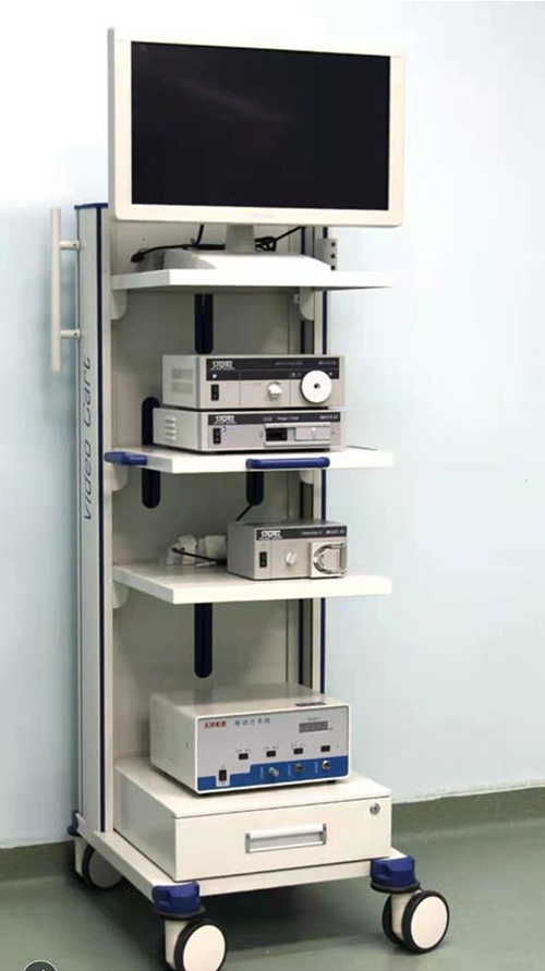
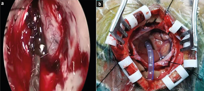
Minimally invasive NES was performed in minimally invasive NES group under compound intravenous anesthesia with tracheal intubation. The neuroendoscopy system (Rudolf, Germany) was used, including a working lens and an observation lens at an angle of 0° and 30°, respectively (Fig. 1). The working lens had three channels integrating flushing, suction and equipment. After CT examination, the skull was drilled at the CT slice with the largest volume of hematoma and the closest position of the center of hematoma to the inner plate of cranial bone. A 3-4 cm-long straight incision was made with the bone hole as the center, and a bone hole with a diameter of 1-1.5 cm was drilled on the skull. After the dura mater was cut, the local cortical brain tissues were subjected to bipolar electrocoagulation for a few millimeters and slightly cut open. The combined neuroendoscopic outer sheath was used for puncture along the direction indicated by CT to establish a minimally invasive endoscopic channel. The surgeon removed intracerebral hematoma through the neuroendoscopic outer sheath under direct vision of the neuroendoscope. After thorough hemostasis was confirmed on the hematoma cavity wall at various angles under the neuroendoscope, the hemostatic gauze was used to cover the hematoma cavity wall through the neuroendoscopic outer sheath under direct vision. After operation, a drainage tube was indwelt outside the hematoma cavity under direct vision of the neuroendoscope generally for 3-14 d. Finally, the neuroendoscopic outer sheath was withdrawn, the bone hole was filled with gelatin sponge, and the skin was sutured (Fig. 2a).


Figure 1. Minimally invasive neuroendoscopic surgery system.


Figure 2. Intervention images of (a) minimally invasive NES and (b) SBWC microsurgery.


Small bone window craniotomy microsurgery


SBWC microsurgery was conducted in SBWC microsurgery group under compound intravenous anesthesia with tracheal intubation. A 6-7 cm-long preauricular oblique incision was made towards the parietal tuber, the temporalis was cut off along the muscle fibers, and the surgical field was expanded with a posterior fossa retractor. The bottom side of bone window (4 cm × 5 cm) should reach the base of the middle cranial fossa as far as possible, the dura mater was cut radially to expose the lateral fissure cistern, and an operating microscope was placed. In the case of high brain pressure and brain tissue prolapse, the brain puncture needle could be punctured into the hematoma cavity through the avascular area in the superior temporal gyrus according to the CT location, so as to aspirate part of the hematoma. After the brain pressure declined, the arachnoid of the lateral fissure cistern was dissected under the microscope, and the insular cortex was exposed. Then the insular cortex was cut open into the hematoma cavity in the avascular area at the slice with the largest volume of hematoma and closest to the insular cortex, and the hematoma was gradually removed under the microscope. The hematoma cavity wall was covered with brain cottons from shallow to deep, and the brain cottons were removed from inside out after the hematoma was removed. The lateral ventricle puncture and external drainage were performed first for patients with hemorrhage breaking into the ventricle (Fig. 2b).

Observation indices


The operation time, intraoperative blood loss and intraoperative hemostasis time were recorded. The difference in the GCS score of patients before operation and at 24 h after operation was detected. The volume of hematoma was calculated using the Tada’s formula based on head CT scan results at 1 d after operation; hematoma clearance rate = (preoperative volume of hematoma – postoperative volume of hematoma)/preoperative volume of hematoma × 100%. Hematoma clearance rate >95% indicated complete clearance, 80-95% indicated nearly complete clearance, and <80% indicated partial clearance. At 72 h after operation, head CT was conducted, and the volume of brain edema zone in the surgical area was compared after the two operations; volume of brain edema zone = total lesion volume on CT scan - volume of hematoma. All patients enrolled were followed up regularly, and the GCS score was given at six months after operation to determine the prognosis: 5 points = good recovery, 4 points = mild disability, 3 points = severe disability, 2 points = vegetative survival, and 1 point = death.

Statistical analysis


SPSS 23.0 software was used for analysis. Normally distributed measurement data were expressed as mean ± standard deviation, and intergroup comparison was conducted by two-sample t test. Numerical data were expressed as percentage, and intergroup comparison was performed by χ2 test. Ranked data were compared by rank sum test. p <0.05 was considered statistically significant.


Results


Operation conditions


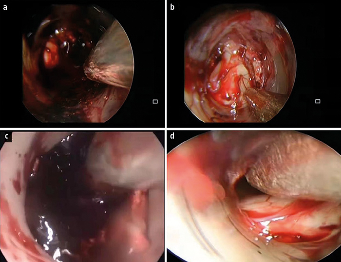
In minimally invasive NES group, the operation time and intraoperative hemostasis time were significantly shorter (127.28 ± 14.21 min and 22.32 ± 2.09 min), and the intraoperative blood loss (43.29 ± 5.46 mL) was significantly less than those in SBWC microsurgery group (193.41 ± 11.56 min and 24.37 ± 2.18 min; 193.28 ± 11.25 mL, p < 0.001). The typical images before and after minimally invasive NES are exhibited in figure 3.


Figure 3. Typical images before and after minimally invasive NES. a and b) images of a 60-year-old male patient before and after hematoma removal; c and d) images of a 56-year-old female patient before and after hematoma removal.


Glasgow coma scale scores


The preoperative GCS score had no significant difference between minimally invasive NES group and SBWC microsurgery group (8.64 ± 1.04 vs. 8.68 ± 1.02 points, p > 0.05). At 24 h after operation, the GCS score in minimally invasive NES group significantly rose (12.89 ± 1.56 points), and it had a significant difference from that in SBWC microsurgery group (11.18±1.14 points, p < 0.001).

Hematoma clearance rates


In minimally invasive NES group, there were 48 cases of complete clearance, 23 cases of nearly complete clearance, 15 cases of partial clearance, and 4 cases of rebleeding. In SBWC microsurgery group, there were 24 cases of complete clearance, 35 cases of nearly complete clearance, 20 cases of partial clearance, and 5 cases of rebleeding. The efficacy had a significant difference between the two groups (p < 0.05). According to the mean rank, minimally invasive NES group had a higher hematoma clearance rate than SBWC microsurgery group (Table II).


Table II. Hematoma clearance rates.
 

SBWC microsurgery group
(n = 84)

Minimally invasive NES group
(n = 90)

z

p


Complete clearance
 

24

48

3.287

0.008


Nearly complete clearance
 

35

23


Partial clearance
 

20

15


Rebleeding
 

5

4


Mean rank
 

40.27

28.65


SBWC: small bone window craniotomy.


Brain edema at 72 h after operation


The volume of brain edema zone in minimally invasive NES group was significantly smaller than that in SBWC microsurgery group (11.82 ± 3.25 mL vs. 18.89 ± 3.15 mL) (p < 0.001).

Prognosis results


A significant difference was found in the distribution of GOS score between the two groups at six months after operation (p < 0.05). According to the mean rank, the prognosis was better in minimally invasive NES group than that in SBWC microsurgery group (Table III).


Table III. GOS scores at six months after operation.
 

SBWC microsurgery group
(n = 84)

Minimally invasive NES group
(n = 90)

z

p


1 point
 

5

6

3.975

0.003


2 points
 

16

6


3 points
 

50

45


4 points
 

10

24


5 points
 

3

9


Mean rank
 

38.92

30.16


SBWC: small bone window craniotomy.


Discussion


The surgical operation of HICH mainly aims to eliminate hematoma as soon as possible, reduce intracranial pressure and alleviate neurological damage caused by brain edema, creating favorable conditions for the functional recovery of damaged brain tissues [8,9]. However, no standardized program has been established on the surgical indications and methods for HICH yet currently, and the selection of treatment methods has always been hotly debated [10,11]. The fundamental objective of surgical treatment of HICH is to ‘strive to save lives and preserve or restore the nerve function to the largest extent’ [12]. With the development of medical technology and the improvement in endoscopic techniques, neuroendoscope has been able to be used alone as a lighting and observation tool like a microscope. The surgeon can perform neurosurgical operations under the neuroendoscope as a ‘handheld microscope’ [13]. Minimally invasive techniques such as neuroendoscopy has gradually attracted the attention of clinicians in the treatment of patients with HICH, but the research is still limited to a small number of case reports, and there are few related studies compared with the multi-index system of the existing mature microsurgery. In this paper, therefore, patients with HICH were treated with minimally invasive NES and SBWC microsurgery, respectively, and the clinical efficacy was compared between the two operation methods, so as to provide references for the selection of operation methods.

In minimally invasive NES group, the bleeding point could be closely observed by the neuroendoscope lens as needed after the hematoma was removed during operation. Under the neuroendoscope, the surgical field could be clearly observed without dead angles, and the hidden bleeding points in the blind zone under the microscope could be found easily (they were mostly lenticulostriate artery small branch bleeding and venous oozing of hematoma cavity wall). The bleeding was stopped effectively by bipolar electrocoagulation or hemostatic materials. Hemostasis was successful in 86 cases and failed in 4 cases, and the hemostatic effect of the latter was satisfactory after prolonging the time of wound compression and spraying biomedical fibrin glue. In SBWC microsurgery group, it was easier to aspirate the hematoma in the surgical channel, but it became difficult to observe directly after the hematoma left the channel for a certain distance, so it was needed to continuously adjust the traction angle of the brain retractor and the light projection direction of the microscope and explore in all directions. Moreover, the lenticulostriate artery branch bleeding in the surgical channel could also be easily found and handled, but there were certain blind zones in the observation of hematoma cavity wall bleeding, easily restricted by the viewing angle. In contrast, the angle of neuroendoscope could be adjusted at any time for close observation. In this study, bleeding was successfully stopped in 70 cases in SBWC microsurgery group. In the remaining 14 cases, the brain tissues were pulled, the microscope angle was adjusted, electrocoagulation was conducted repeatedly, and the wound was compressed by hemostatic materials and covered with biomedical fibrin glue, which was very time-consuming. In some areas with insufficient light projection of the microscope, it was hard to accurately identify the bleeding point, and the hemostatic effect was acceptable. To sum up, the bleeding points can be directly observed in minimally invasive NES, so that the repeated cauterization under poor visibility is avoided in the suspicious area, which can reduce surgical damage and intraoperative blood loss, with relatively simple hemostasis.

SBWC microsurgery can achieve hemostasis under a clear field while removing intracerebral hematoma under the microscope, reducing the risk of rebleeding. After most of the hematomas are removed, however, the brightness of the light source of microscope will gradually decline with the extension of the surgical channel, and there are still blind zones under the microscope even after the light projection angle of the microscope is altered. As a result, it is necessary to pull the brain tissues to remove the hematoma in blind zones, making it difficult to minimize the damage to normal brain tissues. SBWC microsurgery has a higher requirement for micromanipulation, with an emphasis on non-invasive or at least minimally invasive separation of the lateral fissure cistern. The lateral fissure cistern and its arachnoid can be classified into 4 types. It is relatively easy to dissect and separate the first and second types, but it is fairly difficult to separate the third type, and the fourth type belongs to the arachnoid adhesions after meningitis, which can hardly be separated [14]. In such a case, it is time-consuming and inefficient to separate the arachnoid, the risk of damage to the lateral fissure vessels and adjacent brain tissues is extremely high, and cerebral vasospasm occurs easily, thus worsening neurological impairment.

During the evacuation of hematoma, NES is characterized by sufficient illumination and field of view, and it can accurately distinguish the boundary between hematoma and surrounding brain tissues, completely eliminate the hematoma and realize sufficient decompression. In particular, the hematoma cavity in the deep can be observed in NES, and the hematoma in the dead angle under the microscope can also be fully displayed and removed under direct vision. Moreover, intraoperative active bleeding and micro bleeding can be closely observed under the neuroendoscope, which has high imaging quality, high definition and a wide observation angle, greatly reducing the secondary damage to the important nerve tissues around the hematoma during the evacuation of hematoma. Besides, the bleeding can be fully stopped under direct vision, reducing the rebleeding rate after operation. Nishihara et al [15] reported that the average clearance rate was as high as 96% in the emergency treatment of intracranial and intraventricular hematomas with transparent catheter sheath.


Conclusions


In conclusion, NES has the following advantages over SBWC microsurgery in the treatment of HBGH: a) It has mild invasion into brain tissues, and no continuous brain retraction; b) The surgical channel can be observed closely at all corners, and the surgical field is clear; c) It has short operation time, small intraoperative blood loss, and reliable hemostasis. With the continuous development of neuroendoscopic equipment and techniques, the continuous improvement in surgical skills of surgeons, and the accumulation of cases, minimally invasive NES is expected to become an important surgical treatment method for HBGH.


References


 1. Liu J, Li Q, Ren J, Liang X, Zhang Q, Han Y. Association of gender with serum potassium, sodium, and calcium disorders after hypertensive intracerebral hemorrhage. World Neurosurgery 2020; 141: e367-73.

 2. Xu X, Zhang J, Yang K, Wang Q, Chen X, Xu B. Prognostic prediction of hypertensive intracerebral hemorrhage using CT radiomics and machine learning. Brain Behav 2021; 11: e02085.

 3. Shao X, Wang Q, Shen J, Liu J, Chen S, Jiang X. Comparative study of micro-bone window and conventional bone window microsurgery for hypertensive intracerebral hemorrhage. J Craniofac Surg 2020; 31: 1030-3.

 4. Long J, Chen Y, Ming L. Study of correlated factors on prognosis of surgical practices for hypertensive basal ganglia hemorrhage. Chinese Journal of Practical Nervous Diseases 2013; 16: 13-5. 

 5. Yang Z, Zhen Y, Chen K. Clinical effect of hematoma removal with small bone window via lateral fissure island on hypertensive cerebral basal ganglia hemorrhage. Modern Hospitals 2019; 19: 281-3.

 6. Ishikawa T, Ebihara K, Endo K, Endo Y, Sato N, Ota M. Usefulness of a new neuroendoscope brain cylinder for intracerebral hematoma surgery. Interdisciplinary Neurosurgery 2019; 15: 53-9.

 7. Hayashi T, Karibe H, Akamatsu Y, Narisawa A, Shoji T, Sasaki T, et al. Endoscopic hematoma evacuation for intracerebral hemorrhage under local anesthesia: factors that affect the hematoma removal rate. World Neurosurg 2019; 126: e1330-6.

 8. Mindermann T, Heckl S, Mack A. High incidence of transient perifocal edema following upfront radiosurgery for intraventricular meningiomas. Acta Neurochir (Wien) 2020; 162: 2177-82.

 9. Wagner KR, Dwyer BE. Hematoma removal, heme, and heme oxygenase following hemorrhagic stroke. Ann N Y Acad Sci 2004; 1012: 237-51.

 10. Aiyagari V. The clinical management of acute intracerebral hemorrhage. Expert Rev Neurother 2015; 15: 1421-32.

 11. Adeoye O, Ringer A, Hornung R, Khatri P, Zuccarello M, Kleindorfer D. Trends in surgical management and mortality of intracerebral hemorrhage in the United States before and after the STICH trial. Neurocrit Care 2010; 13: 82-6.

 12. Gui C, Gao Y, Hu D, Yang X. Neuroendoscopic minimally invasive surgery and small bone window craniotomy hematoma clearance in the treatment of hypertensive cerebral hemorrhage. Pak J Med Sci 2019; 35: 377.

 13. Sun S, Li Y, Zhang H, Wang X, She L, Yan Z, et al. The effect of mannitol in the early stage of supratentorial hypertensive intracerebral hemorrhage: a systematic review and meta-analysis. World Neurosurg 2019; 124: 386-96.

 14. Chen G, Dong W. Clinical effect comparison of neuroendoscopic minimally invasive surgery and window craniotomy microsurgery in treating hypertensive basal ganglia intracerebral hemorrhage. China Medical Herald 2015; 7: 51-4.

 15. Nishihara T, Nagata K, Tanaka S, Suzuki Y, Izumi M, Mochizuki Y, et al. Newly developed endoscopic instruments for the removal of intracerebral hematoma. Neurocrit Care 2005; 2: 67-74.


Aplicación de la neurocirugía endoscópica en el tratamiento de las hemorragias hipertensivas en los ganglios basales

Introducción. La relevancia de la neurocirugía endoscópica en el tratamiento de las hemorragias hipertensivas de los ganglios basales no se conoce en buena medida.

Objetivo. Comparar la eficacia clínica de la neurocirugía endoscópica mínimamente invasiva con la de la microcirugía con craneotomía de ventana pequeña (SBWC) en el tratamiento de las hemorragias hipertensivas de los ganglios basales.

Pacientes y métodos. Análisis retrospectivo de los datos clínicos de 174 pacientes con hemorragia hipertensiva de los ganglios basales tratados en nuestro hospital desde enero de 2018 hasta septiembre de 2020. Los pacientes se dividieron en dos grupos: uno sometido a neurocirugía endoscópica mínimamente invasiva (n = 90) y otro a microcirugía con SBWC (n = 84). Se compararon la duración de la operación, la tasa de eliminación del hematoma, la recidiva hemorrágica y el pronóstico.

Resultados. En el grupo sometido a la endoscopia mínimamente invasiva, tanto la duración de la intervención como el tiempo de hemostasia fueron significativamente más breves, y la pérdida de sangre durante la intervención fue significativamente menor que en el grupo de microcirugía con SBWC (p < 0,001). La puntuación preoperatoria de la escala de coma de Glasgow (GCS) era de 8,64 ± 1,04 puntos en el grupo de la endoscopia y de 8,68 ± 1,02 puntos en el de la microcirugía (p > 0,05). A las 24 horas de la intervención, la puntuación de la GCS en los sometidos a la neuroendoscopia aumentó hasta 12,89 ± 1,56, con una diferencia significativa respecto al grupo de la microcirugía, que presentaba 11,18 ± 1,14 puntos (p < 0,001). El volumen del edema cerebral fue de 11,82 ± 3,25 mL en el grupo de la neuroendoscopia mínimamente invasiva y de 18,89 ± 3,15 mL en el de la microcirugía (p < 0,001). En comparación con el grupo sometido a esta última, en el grupo de la endoscopia, la eliminación del hematoma fue más extensa y el pronóstico resultó más favorable.

Conclusiones. La neurocirugía endoscópica mínimamente invasiva se mostró más eficaz que la microcirugía con SBWC en el tratamiento de las hemorragias hipertensivas de los ganglios basales.

Palabras clave. Cirugía mínimamente invasiva. Craneotomía. Hemorragia hipertensiva de los ganglios basales. Microcirugía. Neurocirugía endoscópica. Ventana ósea pequeña.


© 2022 Revista de Neurología

Si ya es un usuario registrado en Neurologia, introduzca sus datos de inicio de sesión.


Rellene los campos para registrarse en Neurologia.com y acceder a todos nuestros artículos de forma gratuita
Datos básicos
He leído y acepto la política de privacidad y el aviso legal
Seleccione la casilla si desea recibir el número quincenal de Revista de Neurología por correo electrónico. De forma quincenal se le mandará un correo con los títulos de los artículos publicados en Revista de Neurología.
Seleccione la casilla si desea recibir el boletín semanal de Revista de Neurología por correo electrónico. El boletín semanal es una selección de las noticias publicadas diariamente en Revista de Neurología.
Seleccione la casilla si desea recibir información general de neurologia.com (Entrevistas, nuevos cursos de formación, eventos, etc.)
Datos complementarios

Se os solicita los datos de redes para dar repercusión por estos medios a las publicaciones en las que usted participe.

En cumplimiento de la Ley 34/2002, de 11 de julio, de Servicios de la Sociedad de la Información y de Comercio Electrónico (LSSI-CE), Viguera Editores, S.L.U. se compromete a proteger la privacidad de sus datos personales y a no emplearlos para fines no éticos.

El usuario otorga su consentimiento al tratamiento automatizado de los datos incluidos en el formulario. Los datos facilitados se tratarán siempre con la máxima confidencialidad, salvaguardando su privacidad y con los límites que establecen las leyes vigentes en España, y nunca se cederán a personas ajenas a la organización.

Usted tiene derecho a rectificar sus datos personales en cualquier momento informándolo a secretaria@viguera.com. También se le informa de la posibilidad de ejercitar el derecho de cancelación de los datos personales comunicados.


¡CONVIÉRTASE EN USUARIO PREMIUM DE NEUROLOGIA.COM!

Además, por convertirte en usuario premium, recibirá las siguientes ventajas:

  • Plaza asegurada en todos nuestros Másteres (www.ineurocampus.com)
  • Descuento del 5% en los cursos de “Actualización en Neurología”, la FMC que estará disponible próximamente en la web.
  • Descarga gratuita en formato PDF dos de las obras con más éxito publicadas por Viguera Editores:
    • Oromotors Disorders in childhood (M. Roig-Quilis; L. Pennington)
    • Manual de Neuropsicología 2ª ed. (J. Tirapu-Ustárroz; M. Ríos-Lago; F. Maestú)

El precio para hacerse Premium durante el periodo de un año es de 5€, que podrá pagar a continuación a través de una pasarela de pago seguro con tarjeta de crédito, transferencia bancaria o PayPal:

QUIERO HACERME PREMIUM

No deseo hacerme premium


QUIERO MATRICULARME

No deseo matricularme